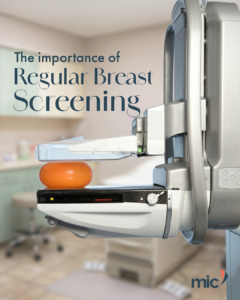
What changes as women age

Not everything about ageing shows on the outside. Your bones, breast tissue and heart all change quietly over time, often without obvious signs. Understanding these changes is the first step to staying proactive about your health. Look out for the next post in our series: What Happens Inside Your Body After 40 or visit our website here to learn more.
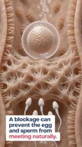

A tiny blockage can make a big difference

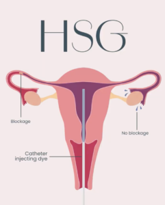
A blocked fallopian tube can quietly stand in the way of pregnancy. The first step is usually an HSG (hysterosalpingogram), which helps confirm whether a blockage is present. If one is found, Fallopian Tube Recanalisation (FTR) is a minimally invasive procedure that may help restore tubal openness in suitable patients. Visit our website to learn more here.

What Happens To Your Bones After Menopause?

Menopause does not just affect how you feel, it can affect your bones too. As oestrogen levels decline, bone loss can happen quickly and often silently, sometimes only becoming clear after a fracture. A DEXA scan helps catch these changes early.

Why dense breast tissue can hide abnormalities

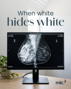
Dense breast tissue can make mammograms more challenging to interpret because both healthy fibroglandular tissue and certain breast abnormalities appear white on X-ray images. This “white-on-white” effect can make small changes more difficult to detect, potentially masking findings that may require further investigation. In addition to affecting image interpretation, dense breast tissue is also recognised as an independent risk factor for breast cancer. Understanding breast density is an important part of breast health, helping guide appropriate screening and, where necessary, additional imaging. To learn more about our Women’s Imaging services, visit our website here.

FAQ: How big do fibroids have to be before treatment?

When it comes to uterine fibroids, size is only one part of the clinical picture. A large fibroid may not require treatment if it remains symptom-free, while a smaller fibroid can significantly affect quality of life depending on its location and the symptoms it produces. Factors such as heavy menstrual bleeding, pelvic pain, pressure on surrounding organs, fertility concerns, and distortion of the uterine cavity all play an important role in determining the most appropriate treatment. Performed through a tiny incision in the groin, Uterine Fibroid Embolisation (UFE) is a minimally invasive treatment that can be considered for women whose fibroids are affecting their quality of life. If you  would like to learn more about UFE, visit our website here

How many samples are taken during a breast biopsy?

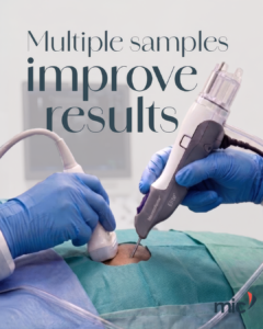
During a vacuum-assisted breast biopsy (VABB), obtaining multiple tissue samples is key to building a complete diagnostic picture. Using a specialised vacuum-assisted device, a radiologist can typically retrieve between 8 and 12 tissue samples through a single, small incision without the need for repeated needle insertions. This technique allows a larger volume of tissue to be collected from the area of concern, improving the quality of the specimen for pathological analysis and iimproving diagnostic confidence. By combining precision imaging with enhanced tissue sampling, VABB plays an important role in the accurate assessment of breast abnormalities. To learn more about Vacuum-Assisted Breast Biopsy, please visit our website here.

What does the dye actually show during an HSG?

Every movement of the contrast dye during a Hysterosalpingogram (HSG) provides valuable diagnostic information. As the dye fills the uterine cavity and travels through the fallopian tubes, it creates a clear outline of the reproductive tract under X-ray guidance. Uninterrupted flow through the tubes, with the dye spilling from the fimbrial ends, indicates that the passage is open. When the dye cannot progress beyond a certain point, it may reveal a blockage that could be contributing to infertility. By visualising the reproductive tract in real time, HSG plays an important role in investigating fertility concerns and guiding appropriate treatment. To learn more about HSG, visit our website here.

Why breast cancer screening doesn’t end with a mammogram

A mammogram is one of the most important tools for the early detection of breast cancer, but it is not always the final step in the diagnostic process. If an area of concern is identified, additional imaging such as ultrasound, MRI, or specialised biopsy procedures may be recommended to gather more information. These investigations help radiologists and referring doctors better understand the nature of a finding and determine the most appropriate next steps. Early detection, combined with improved diagnostic evaluation, plays a vital role in supporting timely treatment and improved patient outcomes. To learn more about women’s imaging services, please visit our website here.

How imaging helps doctors plan treatment, not just make diagnoses

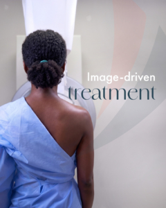
Diagnostic imaging does more than help doctors make a diagnosis. It provides detailed insight into what is happening inside the body, allowing healthcare professionals to assess the location, size, and extent of a condition with greater accuracy. Using advanced technologies such as ultrasound, CT, MRI, and X-ray imaging, doctors can gather valuable information that helps guide treatment decisions and monitor a patient’s progress over time. This detailed understanding supports more precise care, helping to improve outcomes and ensure patients receive the most appropriate treatment for their individual needs. To learn more about our diagnostic imaging services, visit our website here.

The importance of regular breast screening

Interventional Radiology (IR) is a specialised branch of medicine that uses advanced imaging technology to diagnose and treat a wide range of conditions throughout the body. Using guidance from X-ray, ultrasound, CT scans, and other imaging techniques, interventional radiologists can perform targeted procedures through tiny incisions using small instruments such as catheters. Some Interventional Radiology procedures offer a minimally invasive alternative and often resulting in shorter recovery times, less discomfort, and reduced hospital stays. To learn more about Interventional Radiology visit our website here.