Why ports are placed in the chest region?

A chemo port, also known as a port-a-cath, is a small device placed under the skin to allow long-term access to the bloodstream during chemotherapy treatment. It is most commonly positioned in the upper chest because this location provides direct access to large central veins, ensuring safe and efficient delivery of medication, blood samples, and fluids. Placement in the chest also allows the device to remain stable, discreet, and less affected by everyday movement. By using a central vein access point, a chemo port helps reduce repeated needle insertions into smaller veins, preserving vein health and improving overall treatment comfort for patients undergoing long-term therapy 🌿
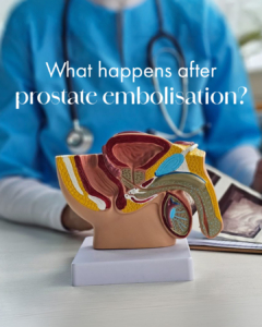
What happens after prostate embolisation?

During Prostate Artery Embolisation (PAE), microscopic particles are used to block the blood supply feeding the enlarged prostate. Once this blood flow is reduced, the prostate begins a gradual process of shrinkage over the following weeks and months. As the tissue decreases in size, pressure on the urethra is relieved, allowing urine to flow more freely. This leads to a progressive improvement in symptoms such as frequent urination, weak stream, and interrupted sleep due to nighttime urination. If you have been diagnosed with an enlarged prostate and would like to explore treatment options, visit our website.

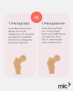
Osteopenia vs Osteoporosis

Bone health is assessed using a DEXA scan, a specialised imaging test that measures bone mineral density using low-dose X-rays. It is used to diagnose conditions such as osteopenia and osteoporosis, and to assess fracture risk over time. Osteopenia refers to a mild reduction in bone density, where bones are weaker than expected but not yet in the osteoporosis range. Osteoporosis is more advanced, where bone density is significantly reduced, increasing the risk of fractures from minor stress or falls. Results are reported using a T-score system, which helps classify bone strength and guide clinical management. Early detection allows for timely intervention to support long-term bone health. To learn more about DEXA scanning, visit our website

A tiny incision with UFE

Uterine Fibroid Embolisation (UFE) is a minimally invasive procedure performed by an interventional radiologist for the management of uterine fibroids. The procedure involves the insertion of a catheter, commonly through a small access point at the groin, using imaging guidance to reach the uterine arteries. Small particles are then introduced to reduce blood flow to selected fibroids. Reducing the blood supply may cause fibroids to decrease in size over time and may help address symptoms associated with fibroids, including heavy menstrual bleeding, pelvic pain and pelvic pressure. Management options for uterine fibroids vary depending on individual clinical circumstances, and patients are advised to discuss appropriate treatment options with their healthcare practitioner. To learn more about UFE, visit our website

FAQ: What’s the difference between a Radiologist and Radiographer?

The terms radiologist and radiographer are sometimes used interchangeably, but they refer to different roles within medical imaging.

A radiographer is a trained healthcare professional who performs imaging examinations such as X-rays, CT scans and MRI scans. Their role includes operating imaging equipment and ensuring images are obtained safely and accurately.

A radiologist is a medical doctor who specialises in the interpretation of medical images. Radiologists analyse imaging studies to assist in diagnosis and may also perform image-guided procedures used in diagnosis and treatment.

In a radiologist-led practice, imaging does not end with image acquisition. It is overseen, interpreted and clinically directed by a specialist doctor from start to finish. To learn more about our services, please visit our website

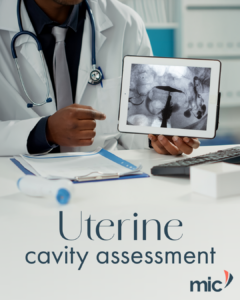
The importance of uterine cavity assessment before FTR

Before Fallopian Tube Recanalisation (FTR) is performed, a detailed assessment of the uterine cavity is essential to ensure the procedure is safe, appropriate, and likely to be effective. This evaluation helps identify intrauterine abnormalities such as adhesions, polyps, or structural variations that may influence fertility or impact treatment planning. If you would like to learn more about FTR or uterine cavity assessment, visit our website.