Imagine a 2026 without fibroids?
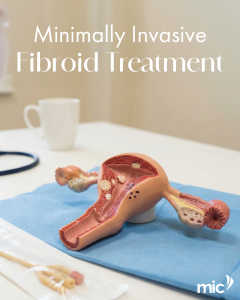
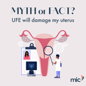
For women diagnosed with symptomatic uterine fibroids, several treatment options exist. One minimally invasive procedure is Uterine Fibroid Embolisation (UFE). UFE is performed by an interventional radiologist. Using image guidance, the specialist accesses an artery through a small puncture (often in the wrist or groin) to deliver tiny particles that reduce the blood supply to the fibroids, which may then shrink over time.
Key Points to Discuss with Your Doctor:
- UFE is a treatment option, not a cure, and its suitability depends on individual factors like fibroid size, number, and location.
- It is considered a uterus-preserving alternative to surgical options.
- A full explanation of potential benefits, risks, and the recovery process should be provided by your gynaecologist and interventional radiologist.
If you have been diagnosed with fibroids, discuss all suitable management options, which may include monitoring, medication, or various procedures, with your gynae to determine the most appropriate option for you. To learn more, please visit our website.