Screening vs Diagnostic Mammography
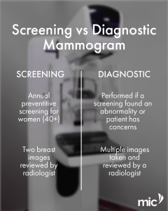
Understanding the difference between screening and diagnostic mammography is essential for effective breast care. Screening mammography is performed routinely in individuals without symptoms to detect early changes before they become noticeable. Diagnostic mammography is used when symptoms are present, or when findings such as microcalcifications or masses require further evaluation. Together, these approaches support timely and accurate assessment. Early detection saves lives, making it important to understand which test is appropriate for your breast health. To learn more about these procedures, speak to your radiologist or visit our website.