80% statistic

Did you know that up to 80% of women with African ancestry will develop fibroids by the age of 50? A diet low in Vitamin D, fruit and green vegetables, as well as exposure to pollutants, can all be triggers for fibroid development. If you have been diagnosed with fibroids and would like to learn more about UFE, please send us a message online or visit our website 👉🏾 https://mic.co.zw/ufe/ 

Prostate Awareness Month

Did you know that prostate cancer is the second most common cancer among men globally? Fortunately with an early diagnosis, this disease is often curable. If you are a male over 40, your doctor might recommend a PSA blood test for prostate screening. This is typically necessary every two years, depending on your doctor’s recommendation. We hope Prostate Awareness Month inspires you to create this routine if you haven’t already ❤️

Why fibroids cause painful periods

Fibroids are benign tumours in the uterus that can significantly contribute to painful periods. One reason for this is the elevated levels of prostaglandins, naturally occurring chemicals in the body that promote inflammation and pain. When you have fibroids, these prostaglandins can lead to increased inflammation in the uterus and heavier menstrual bleeding. Uterine Fibroid Embolisation (UFE) is a minimally invasive procedure designed to treat fibroids by blocking the blood vessels that supply them. This effectively shrinks the fibroids and alleviates symptoms. If you have been diagnosed with fibroids and would like to learn more about UFE, please send us a message online or visit our website  

FAQ: Can fibroids grow back after UFE?

The answer is yes, but the likelihood is low. Studies show that Uterine Fibroid Embolisation (UFE) effectively treats fibroids, with just a small chance of the targeted fibroids regrowing. It may be the unlikely case that new fibroids form, rather than the recurrence of treated ones. For more information about fibroids and non-invasive solutions, please send us a message or visit our website  

What scans should I schedule?

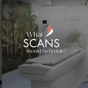
When it comes to booking health scans the type you need will depend on your symptoms and your doctor’s recommendations. Each scan has a unique role in diagnosing and monitoring various conditions👇🏾

➡️ MRI (Magnetic Resonance Imaging): Ideal for soft tissue evaluation, including the brain, spine, joints, and organs.

➡️ CT Scan (Computed Tomography): Provides detailed images of bones, blood vessels, and soft tissues, often used for trauma, cancer, and infection detection.

➡️ Ultrasound: Commonly used for monitoring pregnancy, but also helpful for examining organs like the liver, kidneys, and heart.

➡️ 3D Mammography: Used for early detection of breast cancer by providing detailed images of breast tissue.

For more details on the types of scans we offer and how they can assist you, please send us a message or visit our website 

The 3 types of blocked fallopian tubes

Fallopian tube blockages can occur in different parts of the tube, affecting a woman’s ability to conceive. Blockages can prevent eggs from reaching the uterus or sperm from reaching the egg, leading to fertility issues. The location of the blockage is crucial in determining both the cause and the treatment 👇🏾

✴️ Proximal: Near the uterus, where the fallopian tube begins. 

✴️ Medial: Found in the middle section of the tube, often caused by scarring or inflammation. 

✴️ Distal: Located near the end of the tube, close to the ovaries and fimbriae.

If you have been diagnosed and would like to learn more about blocked fallopian tubes, please send us a message online or visit our website  

Myth or Fact: Only older women get breast cancer

Myth busted! ❌ Breast cancer doesn’t discriminate based on age. In Zimbabwe, the peak age for diagnosis is actually between 40 and 50. No matter your age, it’s important to be aware of the signs and symptoms of breast cancer and schedule regular check-ups. Here’s a quick guide to recommended screenings 👇🏾

🎀25-34 years: Clinical Breast Exam by a nurse or doctor (CBE) every 1-3 years. 

🎀35-39 years: CBE and Ultrasound OR mammography* every 1-3 years.

🎀40-74 years: CBE + Mammography annually.

🎀75 years and older: Discuss with your doctor.

*Mammography may be recommended for women with a higher risk of breast cancer (affected first degree relatives e.g. mother or sister, previous abnormal breast biopsy, previous chest wall radiotherapy, previous breast cancer).

Remember, early detection is key. Let’s prioritise our health!

Why should I test bone density?

As we age, our bones naturally lose mass, which can lead to osteoporosis — a condition where bones become fragile and more likely to fracture. Testing bone density is essential for detecting early signs of bone loss, particularly in women, who are at higher risk as they age. A bone density test or DEXA scan, helps assess the strength and health of your bones, allowing for early intervention and treatment if needed. To learn more about bone density testing please send us a message online or visit our website  

How is the FTR Procedure Performed?


Fallopian Tube Recanalisation (FTR) is a minimally invasive procedure designed to clear blockages in the fallopian tubes, to improve the likelihood of pregnancy. The procedure follows these 4 steps 👇🏾

1️⃣ An intravenous drip is placed and short acting medication is given for sedation. 

2️⃣ A speculum is then placed in the vagina and a catheter is inserted through the cervix into the uterus. 

3️⃣ A liquid dye called ‘contrast’ is injected through a catheter to fill and examine the uterine cavity and fallopian tubes.

4️⃣ When the blockage is found, a small, soft microwire is threaded through the fallopian tube to clear the blockage.

If you have been diagnosed with blocked fallopian tubes and would like to learn more, please send us a message online or visit our website  

What does Gynaecological Imaging do?

Gynaecological imaging plays a vital role in understanding women’s reproductive health. These studies include transabdominal and transvaginal pelvic ultrasounds, which provide detailed views of reproductive organs. There are also pelvic floor ultrasounds to evaluate urinary incontinence and prolapse, along with specialised deep endometriosis ultrasounds. These imaging methods are crucial for diagnosing various conditions, ensuring women receive accurate assessments and tailored treatment options for optimal reproductive health. If you would like to learn more about gynaecological imaging please send us a message online or visit our website