How ultrasound pinpoints exact biopsy locations

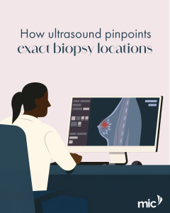
Ultrasound-guided biopsies are highly effective because imaging and needle movement occur simultaneously. As the probe glides over the skin, it produces clear visualisation of the lesion, blood vessels and surrounding anatomy. The radiologist follows these images to ensure the needle reaches the exact location required for sampling. This method improves safety and accuracy, particularly for abnormalities not visible on mammography. It also offers a comfortable, minimally invasive patient experience. For more information about this technique, visit our website.

We’re Open

Season’s greetings from MIC Radiology Group 🌸 We’ll be operating as usual throughout the festive season to ensure continuous service.

Stereotactic Breast Biopsy – What to expect during the procedure.

During a stereotactic breast biopsy, digital mammography guides the radiologist to the precise area requiring assessment. Once the breast is positioned, local anaesthesia is used to numb the region, followed by a small opening that allows the needle to collect tissue samples for diagnosis. This targeted approach minimises trauma and supports a quicker recovery — with most patients able to resume light activities shortly after the procedure. To learn more about this technique,visit our website.

Ideal candidates for TACE

Trans-Arterial Chemoembolisation (TACE) is a minimally invasive alternative for liver cancer patients who are not candidates for surgery. This advanced procedure is ideal for individuals with tumours confined to the liver and good overall health. It works with precision — delivering chemotherapy directly to the tumour while blocking its blood supply. This dual attack is highly effective for hepatocellular carcinoma and can shrink a tumour, potentially creating a path to future surgical removal.

Understanding PT Stent Insertion

Percutaneous transluminal angioplasty (PTA) or stent insertion is a minimally invasive procedure used to open narrowed or blocked arteries. It is used to restore blood flow and treat conditions like atherosclerosis in various parts of the body, including the legs, kidneys, and heart (in which case it is also called coronary angioplasty). Using real-time imaging, a catheter is guided through a small incision in the wrist or groin to the affected artery. A balloon is then inflated to widen the vessel, followed by the placement of a small metal stent to keep the artery open and ensure continuous blood flow.

How chemo ports improve long-term chemotherapy management

For patients undergoing prolonged chemotherapy, a chemo port (or port-a-cath) can help to simplify treatment. This small, implantable device is placed under the skin, typically in the upper chest, and provides a reliable access point to a major vein for administering drugs, drawing blood, and delivering nutrients. It helps to preserve vein health by reducing the need for repeated needle sticks. While ports can make treatment more comfortable, it is important to discuss both the benefits and potential risks with your doctor. The decision to use a chemo port is made by your oncologist or treating physician.

Understanding PAE’s impact on overall prostate health

Prostate Artery Embolisation (PAE) is a minimally invasive radiological procedure that uses tiny particles to reduce blood flow to the prostate. It’s performed through a small access point in the wrist or groin under image guidance. In appropriately selected patients, reduced blood flow can lead to gradual prostate size reduction in patients with benign prostatic hyperplasia (BPH). This results in possible improvement in urinary symptoms over weeks to months and typically shorter recovery than open surgery (varies by individual). Suitability is assessed by your doctor considering symptoms, prostate size and overall health. Alternatives such as medication, watchful waiting or surgery such as TURP/laser may be recommended. If you’ve been diagnosed with BPH, speak to your doctor about PAE as a treatment option, or visit our website to learn more.

How Micro Particles Reduce Prostate Size

During a Prostate Artery Embolisation (PAE) procedure, our specialists use microscopic particles to reduce blood flow in the arteries supplying the prostate. This minimally invasive procedure is performed through a small access point in the wrist or groin.

The goal of PAE is to achieve gradual shrinkage of the prostate for symptom relief, while aiming to preserve surrounding healthy structures. If you’ve been diagnosed with BPH, speak to your doctor about PAE as a treatment option, or visit our website to learn more.

Step by Step Overview of PAE

Prostate Artery Embolisation (PAE) is a minimally invasive procedure that may be an option for treating Benign Prostatic Hyperplasia (BPH).
Here is an overview of how the procedure is typically performed 👇🏾
1️⃣ Advanced imaging is used to map the arteries supplying the prostate.
2️⃣ A catheter is gently inserted through a tiny incision in the wrist or groin.
3️⃣ Microscopic particles are deployed to reduce blood flow to the targeted areas.
4️⃣ The intended outcome is for the prostate to gradually shrink, which aims to relieve urinary symptoms.

If you’ve been diagnosed with BPH, speak to your doctor about PAE as a treatment option, or visit our website to learn more.

Men’s Health Month

This Men’s Health Month, we are highlighting the importance of proactive screening and early intervention for prostate and testicular cancer. Early prostate and testicular cancer may have minimal symptoms, that’s why it’s important that men talk to their GP about their prostate and testicular health and the available tests. Radiology, including minimally invasive procedures play a key role in early diagnosis. This November, take a proactive step — talk to your doctor about prostate and testicular cancer screening.