What happens during PT Stent Insertion

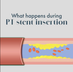
During a stenting procedure, flexible catheters and a balloon are used to reopen a blocked artery. The catheter with an uninflated balloon is guided to the site of the obstruction, then the balloon is inflated to widen the vessel. Once the artery is expanded, a permanent mesh stent is placed to maintain consistent blood flow. If multiple blockages are present, the procedure may be repeated to support comprehensive cardiovascular health. Patients may feel slight pressure at the insertion site or mild discomfort during inflation, but the technique is designed to be as comfortable as possible ⚕️

How Ultrasound-Guided Biopsies are performed

When a mammogram or physical examination reveals a suspicious mass, an ultrasound-guided breast biopsy provides a precise, minimally invasive method to obtain tissue samples. High-frequency sound waves visualise the lesion in real time, allowing the radiologist to position the needle  without major surgery. After local anaesthesia numbs the area, a tiny incision is made to extract multiple samples for microscopic evaluation. This technique avoids ionising radiation, leaves minimal scarring, and is usually performed on an outpatient basis, with most individuals returning home shortly after. To learn more about how this diagnostic tool visit our website.

ISO Certification

MIC Radiology Group has received ISO 9001:2015 certification following an independent audit of its Quality Management System. This certification relates to the organisation’s quality management processes and reflects compliance with the requirements of the ISO 9001:2015 standard.

The certificates were formally handed over by the Director General of the Standards Association of Zimbabwe, Mr Cosmus Mukoyi, during the handover ceremony.

The Quality Management System includes structured processes relating to areas such as patient scheduling, imaging workflows, reporting processes, and quality assurance. MIC Radiology Group remains committed to maintaining ISO Quality standards.

Symptom relief with PAE

An enlarged prostate can cause distressing urinary symptoms, including frequent night-time voids, weak urine flow, and urgency. Prostate Artery Embolisation (PAE) is a minimally invasive procedure that reduces blood flow to the prostate, causing it to shrink and relieving pressure on the urethra. This targeted approach improves urine flow, reduces urgency, and minimises night-time interruptions, while carrying a lower risk of side effects compared to traditional surgery. Patients often experience faster recovery and a swift return to daily activities. To learn more about PAE, visit our website.

Does hormonal imbalance affect fertility?

Hormonal irregularities and physical tubal blockages represent two distinct challenges. While procedures such as Fallopian Tube Recanalisation may address mechanical obstructions, hormonal imbalances can influence fertility by affecting the regularity of ovulation.Fluctuations in chemical messengers, including Follicle‑Stimulating Hormone and Luteinising Hormone, may interfere with the release or maturation of an egg.Achieving a successful pregnancy often requires a comprehensive assessment to ensure both structural pathways are clear and the endocrine system is functioning optimally 🌸

Mapping the GI Tract

Diagnostic examinations of the digestive tract often utilise contrast agents, such as barium, to enhance internal visibility during X-ray or fluoroscopic procedures. These substances coat the mucosal lining of the oesophagus, stomach, and intestines, allowing for a detailed evaluation of their internal structure and function. By creating a clear silhouette of the GI tract, this technique helps identify conditions such as strictures, ulcers, or abnormal motility. Such precise imaging is essential for achieving improved diagnosis and formulating an effective treatment plan. If you would like to learn more about gastrointestinal imaging, visit our website.

Mapping abnormalities with Stereotactic precision

Stereotactic breast biopsy is an advanced, minimally invasive procedure used to sample abnormalities that may not be visible on a standard ultrasound. A specialised digital mammography machine converts x-rays into electronic signals to produce high-resolution images. By taking images from two angles, the computer calculates the exact coordinates of the target, creating a precise map of microcalcifications or small lesions. A radiologist then inserts a needle through a small incision to collect tissue samples with minimal disruption to surrounding tissue. This technique improves accurate diagnosis and supports early detection and management of non-palpable breast lesions. To learn more, visit our website.

Happy International Women’s Day!

Today we celebrate the strength, resilience and accomplishments of women around the world 🌸 Happy International Women’s Day!

Can UFE be performed multiples times?

Yes in certain cases, Uterine Fibroid Embolisation (UFE) can be performed more than once. UFE works by blocking the blood supply to existing fibroids, causing them to shrink. However, it does not prevent new fibroids from developing in the future. If new fibroids grow and begin causing symptoms, a repeat procedure may be considered. That said, needing a second UFE is uncommon. Most women experience long-term relief after a single treatment. If you have been diagnosed and would like to learn more about UFE, visit our website.

Symptom relief: From neck pressure to swallowing discomfort.

Thyroid Microwave Ablation (MWA) is a minimally invasive treatment for benign thyroid nodules that cause symptoms such as neck pressure, a sensation of fullness in the throat, difficulty swallowing and visible swelling. Using imaging guidance, a fine probe is inserted into the nodule and controlled microwave energy is applied, causing the tissue to shrink gradually over time. As the nodule reduces in size, compression eases and many patients experience meaningful symptom relief, often returning to normal activities quickly with minimal discomfort and no surgical scarring. The procedure is guided by interventional radiologists or endocrinologists, in collaboration with your doctor, to ensure precision and safety. To learn more about whether MWA is suitable for you, speak to your endocrinologist or visit our website.