How (VAB) reduces the need for surgical biopsies.

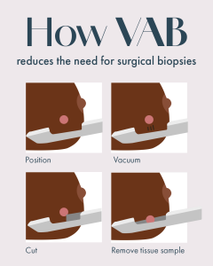
When a breast imaging scan identifies an area requiring further assessment, your doctor may discuss biopsy options. One advanced technique is Vacuum-Assisted Biopsy (VAB). VAB is a minimally invasive, image-guided procedure. A needle attached to a vacuum device is used to collect multiple tissue samples through one small incision. This method allows for the removal of a larger sample of tissue than a traditional core needle biopsy, which can be necessary for a definitive diagnosis in certain situations.The decision to recommend a VAB is made by your doctor and radiologist based on your specific clinical needs 🩺 Speak to your doctor or visit our website for more info.

Imagine a 2026 without fibroids?

For women diagnosed with symptomatic uterine fibroids, several treatment options exist. One minimally invasive procedure is Uterine Fibroid Embolisation (UFE). UFE is performed by an interventional radiologist. Using image guidance, the specialist accesses an artery through a small puncture (often in the wrist or groin) to deliver tiny particles that reduce the blood supply to the fibroids, which may then shrink over time.

Key Points to Discuss with Your Doctor:

  • UFE is a treatment option, not a cure, and its suitability depends on individual factors like fibroid size, number, and location.
  • It is considered a uterus-preserving alternative to surgical options.
  • A full explanation of potential benefits, risks, and the recovery process should be provided by your gynaecologist and interventional radiologist.

If you have been diagnosed with fibroids, discuss all suitable management options, which may include monitoring, medication, or various procedures, with your gynae to determine the most appropriate option for you. To learn more, please visit our website.

Gastrointestinal Imaging

Gastrointestinal imaging helps your doctor visualise your digestive tract. Different tests are used to evaluate different areas and symptoms.

Common Studies Include:

🩺 Contrast Studies (Fluoroscopy):

  • Barium Swallow: Examines the esophagus and stomach.
  • Small Bowel Follow-Through: Tracks the journey of contrast through the small intestine.
  • Enema (Barium or Contrast): Evaluates the large intestine/colon.

🔍 Advanced Cross-Sectional Imaging:

  • CT Colonography: A CT scan that provides detailed 3D images of the colon, often used for polyp detection.
  • MR Enterography: An MRI scan specifically optimized to evaluate the small intestine, excellent for assessing inflammation (e.g., Crohn’s disease).
  • CT Enterography: A CT scan providing detailed images of the small bowel wall and surrounding structures.

These are diagnostic tools, not treatments. The choice of scan—whether a simple contrast study or a complex CT/MR—is made by your referring doctor and the radiologist doctor based on your specific symptoms, medical history, and the clinical question that needs answering.If you have ongoing digestive issues, the first step is a consultation with your healthcare provider to determine the most appropriate diagnostic path for you.

PAE vs. TURP

For men with symptoms of an enlarged prostate (Benign Prostatic Hyperplasia or BPH), understanding different treatment approaches is key.

Two common procedures are:

▪ TURP (Transurethral Resection of the Prostate): A surgical procedure to remove prostate tissue. It is performed under anesthesia and may involve a hospital stay.

▪ PAE (Prostate Artery Embolisation): A minimally invasive, image-guided treatment that reduces blood flow to the prostate, leading to a gradual size reduction.

These treatments differ in technique, invasiveness, and recovery. The choice between them is a medical decision made by your urologist and interventional radiologist based on your specific diagnosis and health profile.

If you have BPH, discuss all suitable management options with your healthcare provider. 

Understanding Your Heart: A Look at Common Cardiac Imaging Studies

Cardiac imaging helps your doctor assess heart structure, function, and blood flow. Different tests provide different information:

❤️ Echocardiogram (Echo): Uses ultrasound waves to create real-time moving images of your heart’s chambers, valves, and pumping action.

❤️ CT Calcium Score: A quick, non-contrast CT scan that measures calcium plaque buildup in coronary arteries, helping assess overall heart attack risk.

❤️ CT Coronary Angiogram: An advanced CT scan using contrast dye to create detailed 3D images of the coronary arteries to evaluate for blockages or narrowing.

❤️ Cardiac MRI: Uses a magnetic field and radio waves to produce highly detailed images of heart muscle, structure, tissue health, and function, without radiation.

These are diagnostic tools. Your cardiologist or doctor will determine if and which test is appropriate based on your individual symptoms, history, and risk factors.

If you have concerns about heart health, the first and most important step is a consultation with your healthcare provider. For more information, visit our website.

CT or MRI Scan: Which is Better?

When your doctor recommends a diagnostic scan, you may wonder about the difference between a CT (Computed Tomography) scan and an MRI (Magnetic Resonance Imaging) scan. Both are powerful imaging tools, but they work differently and provide unique information. CT Scan: Uses X-rays to create detailed cross-sectional images of the body. It is exceptionally fast and excellent for visualising bones, detecting bleeding, and assessing trauma or lung conditions. MRI Scan: Uses a strong magnetic field and radio waves (no ionising radiation) to generate detailed images of soft tissues. It excels at visualising the brain, spinal cord, muscles, ligaments, and organs. The choice between CT and MRI is not about which technology is “better,” but about which is the most appropriate for your specific clinical situation. Your doctor and the radiologist will decide which based on what part of the body needs examination, the suspected condition, and your individual health profile. Visit our website to learn more 👉🏾 mic.co.zw

Your first health check in 2026

This January, as we reset our priorities for the new year, let’s champion our health. It’s Cervical Cancer Awareness Month. Cervical Cancer is the most common cancer in Zimbabwe. Proactive health checks, like cervical cancer screening, play a critical role in early detection, as many conditions present without early symptoms. Make 2026 the year you speak to your doctor or clinic about preventive healthcare. Your health is your priority. 🩺